Expert Opinion

The Need for Separate Testing with Acetylcholine for the Assessment of Endothelial Dysfunction and Coronary Artery Spasm

Register or Login to View PDF Permissions
Permissions× For commercial reprint enquiries please contact Springer Healthcare: ReprintsWarehouse@springernature.com.

For permissions and non-commercial reprint enquiries, please visit Copyright.com to start a request.

For author reprints, please email rob.barclay@radcliffe-group.com.
Information image
Average (ratings)
No ratings
Your rating

Abstract

Intracoronary acetylcholine (ACH) testing is clinically useful to diagnose the presence of the coronary vasomotor disorders coronary endothelial dysfunction and coronary epicardial/microvascular spasm. In Western countries, continuous intracoronary injection of ACH for 2–3 minutes without a pacemaker is the usual method, while rapid injection of ACH for 20–30 seconds with a pacemaker is the traditional procedure in Japan. Coronary microvascular spasm is often observed in Western populations, whereas coronary epicardial spasm is frequently seen in Japanese subjects. Methodological differences between Western and Japanese protocols may lead to the opposite prevalence of coronary vasomotor disorders. This article discusses the optimal method for diagnosing endothelial dysfunction and epicardial/microvascular spasm based on previous reports, and compares intracoronary ACH testing performed by Western cardiologists with that by Japanese physicians.

Disclosure:The authors have no conflicts of interest to declare.

Received:

Accepted:

Published online:

Correspondence Details:Shozo Sueda, Department of Cardiology, Ehime Prefectural Niihama Hospital, Hongou 3 choume 1-1, Niihama City, Ehime Prefecture 792-0042, Japan. E: sue23sho@gmail.com

Open Access:

This work is open access under the CC-BY-NC 4.0 License which allows users to copy, redistribute and make derivative works for non-commercial purposes, provided the original work is cited correctly.

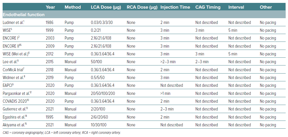
Intracoronary administration of acetylcholine (ACH) was found to induce paradoxical coronary vasoconstriction due to coronary endothelial dysfunction, according to Ludmer et al. in 1986.1 They carried out incremental dose-up injection of ACH (0.03 μg, 0.3 μg, 3 μg and 30 μg) for 2 minutes with an infusion pump into the left coronary artery (LCA). In 2003, the ENCORE study used intracoronary ACH (2.16 μg, 21.6 μg and 108 μg) with an infusion pump via microcatheter for 3 minutes into the LCA to verify the presence or absence of coronary endothelial dysfunction.2 In contrast, in 1986 Yasue et al. reported the usefulness of intracoronary ACH testing in patients with variant angina.3 They used incremental dose-up of ACH (20 μg, 50 μg and 100 μg into the LCA and 20 μg and 50 μg into the right coronary artery [RCA]) for 20–30 seconds with manual injection. The sensitivity and specificity of ACH testing for patients with variant angina were found to be acceptable.3 Intracoronary ACH testing is clinically used for the investigation of coronary endothelial dysfunction and coronary spasm; however, the procedures and doses involved vary worldwide. In this article, we summarise the information on intracoronary ACH testing with the aim of determining the optimal ACH procedure for verifying the presence or absence of coronary endothelial dysfunction and coronary spasm.

Vasoreactivity Testing for Coronary Endothelial Dysfunction

Furchgott and Zawadzki reported on the essential role of endothelial cells in the relaxation of arterial smooth muscle by ACH in vitro.4 If coronary arteries have normal endothelial function without any atherosclerotic lesions, intracoronary injection of ACH dilates the coronary artery. However, if coronary arteries have abnormal endothelial function due to coronary atherosclerosis, intracoronary injection of ACH constricts the coronary artery. As shown in Table 1, the majority of researchers have used relatively low doses of ACH (0.36 μg, 3.6 μg and 36 μg) for 2–3 minutes.5–10 Intracoronary ACH is injected at a constant flow rate using pump infusion without a pacemaker, and the target coronary artery is usually the left anterior descending artery.11–13 In Japan, there are few reports on coronary endothelial dysfunction.14,15

Table 1: Intracoronary Acetylcholine Testing for Coronary Endothelial Dysfunction

Article image

Inconsistency of Intracoronary ACH Testing in the Clinic

Physicians use intracoronary ACH testing to verify the presence of coronary spasm or coronary endothelial dysfunction. Low doses of ACH for 2–3 minutes’ continuous injection are used for the diagnosis of coronary endothelial function, while bolus injection of high-dose ACH for 20–30 seconds is used to investigate the presence of coronary epicardial spasm/coronary microvascular spasm.16 However, physicians may perform additional ACH testing arbitrarily (Tables 1–3). Furthermore, the majority of physicians do not perform vasoreactivity testing on both coronary arteries.7,16 They perform vasoreactivity testing mainly into the left anterior descending artery. If they obtain negative results for intracoronary ACH testing in the LCA, they then carry out intracoronary injection of ACH into the RCA whenever possible.

Duration of ACH Injection: 20 Seconds versus 180 Seconds

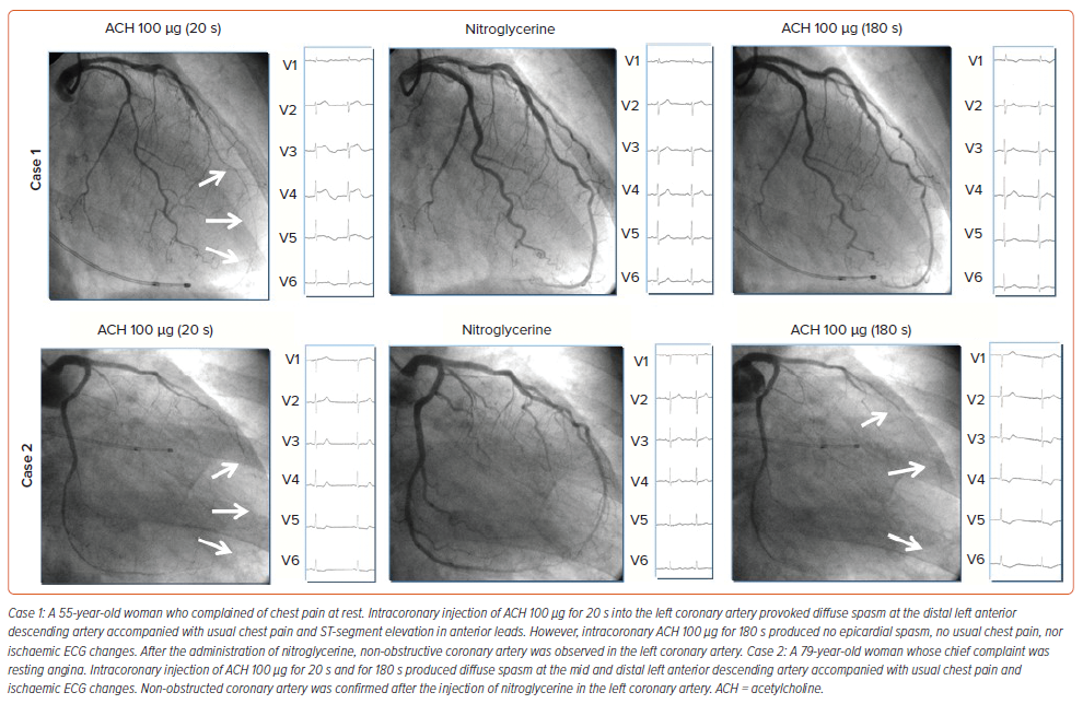
The duration of ACH injection plays an important role in the type of spasm provoked. Given the same doses of ACH in the same patients, rapid injection of ACH provoked more cases of epicardial spasm than moderate administration of ACH.17 Provoked spasm incidence by a 20-second injection of ACH was significantly higher than that by a 180-second ACH injection (73.3%, 22/30 versus 33.3%, 10/30, p<0.05).17 As shown in Figure 1, intracoronary injection of ACH for 20 seconds provoked typical epicardial spasm but that for 180 seconds did not (case 1), whereas in case 2, intracoronary injection of ACH for both 20 seconds and for 180 seconds produced the same coronary responses. Cardiologists should understand the clinical differences underlying the coronary responses between the two procedures, even in the same patients.

Figure 1: Vasoreactivity Testing: Acetylcholine Injection of 20 s versus 180 s

Article image

Transition for Ideal Vasoreactivity Testing of ACH

In 2014, Ong et al. reported on an incremental dose-up of 2 μg, 20 μg, 100 μg and 200 μg ACH manually infused over a period of 3 minutes into the LCA via angiographic catheter.18 The ACH doses in their protocol were derived from the multicentre ENCORE study (Supplementary Figure 1).2 In the ENCORE study, the dose for the left anterior descending artery and for the left circumflex artery was 100 μg into each vessel injected via selective catheter. For practical reasons, in the Ong et al. study the ACH injection was performed unselectively via the diagnostic catheter into the LCA with a maximum dose of 200 μg. In patients who remained asymptomatic and had no diagnostic ST-segment changes during LCA ACH infusion, 80 μg ACH was injected into the RCA.18 In the CASPAR study in 2008, incremental doses of 2 μg, 20 μg and 100 μg ACH were injected into the LCA via the diagnostic catheter for 3 minutes each.19 However, in 2016, Ong et al. reported the use of the same ACH doses with an injection duration of 20 seconds instead of 3 minutes without a pacemaker.20 The maximum ACH dose and the injection time in their study varied from 100 μg ACH to 200 μg ACH and from 3 minutes to 20 seconds, respectively.

In contrast, we performed intracoronary ACH testing from 1991 based on the Japanese Circulation Society (JCS) guidelines.21 Incremental dose-up of 20 μg and 50 μg into the RCA and of 20 μg, 50 μg and 100 μg into the LCA was injected for 20 seconds with a pacemaker. We used a maximum ACH dose of 80 μg into the RCA from 1993, and a maximum ACH dose of 200 μg into the LCA from 2012.22

We have been performing intracoronary ACH testing for more than 30 years, but currently, there is no established global standard for ACH testing. Therefore, an important goal for the future is to establish optimum, unified recommendations for ACH testing for coronary spasm and coronary endothelial dysfunction.

Vasoreactivity Testing for Coronary Epicardial Spasm

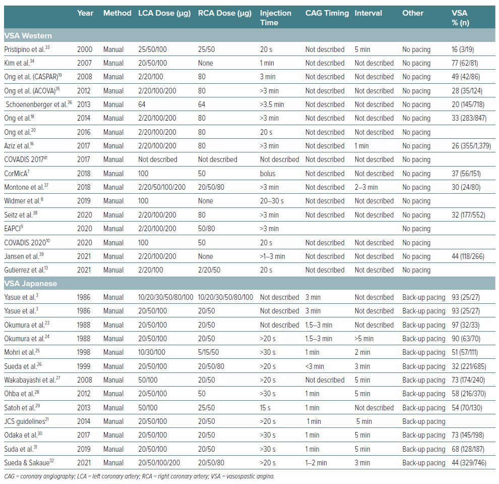
The intracoronary injection time for ACH in Japanese reports is less than 30 seconds, while in the majority of Western reports it is over 3 minutes (Table 2).23–39 Furthermore, back-up pacing was necessary in Japanese reports, whereas in Western reports there was no back-up pacing during vasoreactivity testing of ACH. There are obvious methodological differences between Japanese and Western studies, and hence a unified method is needed to verify the presence or absence of coronary epicardial spasm. In the Western reports, if a positive epicardial spasm was not provoked in the LCA, intracoronary ACH was administered into the RCA. The main target coronary artery is the LCA for Western cardiologists, while Japanese cardiologists perform tests for both coronary arteries if possible. The definition of positive epicardial spasm was similar between Western and Japanese reports.

Table 2: Intracoronary Acetylcholine Testing for Vasospastic Angina

Article image

Vasoreactivity Testing for Coronary Microvascular Spasm

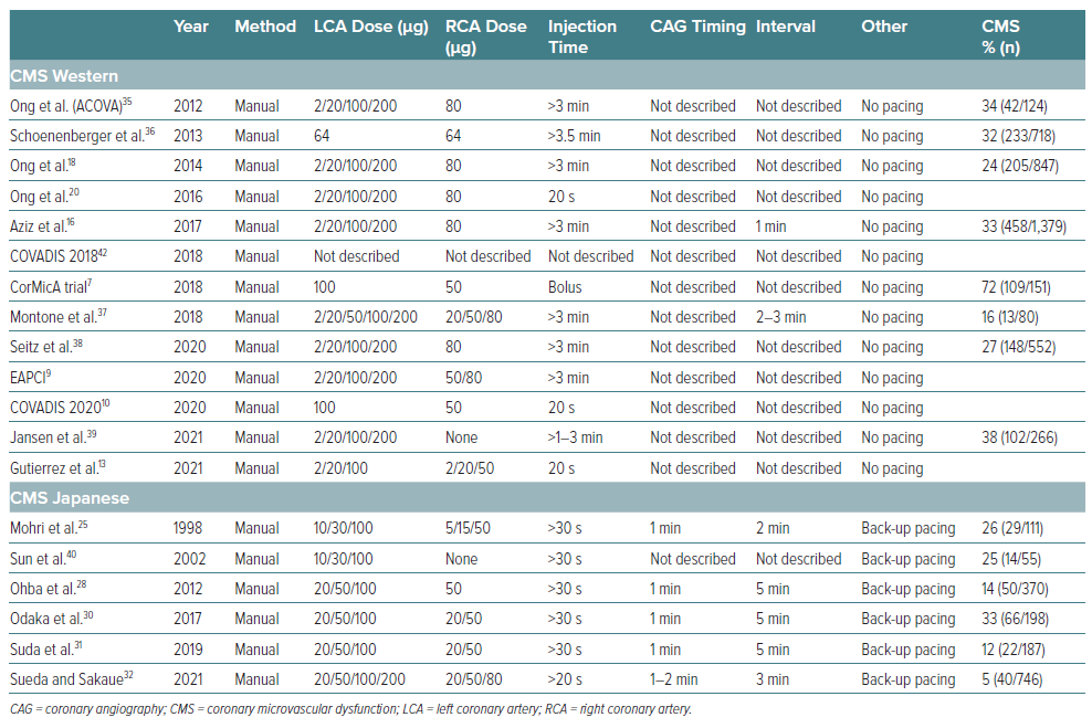
As shown in Table 3, the majority of researchers used the same methods to diagnose the presence or absence of epicardial spasm and coronary microvascular spasm.40 There are no recommendations regarding methodological issues in the COVADIS group 2017/18 reports.41,42 However, COVADIS reports in 2020 provided recommendations on ACH doses for the first time.10 They recommended intracoronary injection of ACH 100 μg into the LCA and ACH 50 μg into the RCA for 20 seconds without a pacemaker. Compared with Western reports, the ACH intracoronary injection time is remarkably short in Japanese reports. Back-up pacemaker insertion is necessary for ACH testing in Japan, while we could not find any mention of the procedure under pacemaker in Western reports.43 The definition of positive coronary microvascular spasm in Western reports is not different from that in Japanese reports.

Table 3: Intracoronary Acetylcholine Testing for Coronary Microvascular Spasm

Article image

Initial Examination: Physiological Functional Tests versus Vasoreactivity Testing

Western guidelines recommend coronary physiological functional measurements before vasoreactivity testing.9 However, Japanese physicians recommend vasoreactivity testing first, before coronary physiological functional measurements.43 Furthermore, Western cardiologists reported that the effect of intracoronary injection of <200 μg nitroglycerine had almost completely disappeared 10 minutes later, possibly due to the short half-life.44 If ACH testing is performed first, then resting flow and coronary flow reserve assessment may be inaccurate, particularly after a positive vasospasm test.17 However, JCS guidelines recommend at least 48 hours’ cessation of coronary vasodilators before coronary vasoreactivity testing. Western guidelines place considerable weight on the identification of coronary microvascular dysfunction, whereas Japanese researchers place importance on diagnosing the presence of epicardial spasm.

These methodological and ethnic differences may be contributing to the disparity in diagnostic strategies. Therefore, if the aim is to diagnose coronary epicardial spasm or coronary microvascular spasm, we suggest that ACH vasoreactivity testing be done first, before the assessment of coronary physiological functioning.

Need for Standardised Vasoreactivity Testing for Coronary Epicardial and Coronary Microvascular Spasm

Although there are ethnic and racial differences in epicardial spasm and coronary microvascular spasm, unified diagnostic testing worldwide is essential for patients with coronary vasomotor disorders, especially given that the definition of positive coronary microvascular spasm and of coronary microvascular dysfunction is not different between Western and Japanese reports. The challenge is, therefore, to establish a unified procedure for diagnosing epicardial spasm and coronary microvascular spasm.

Complications during Intracoronary ACH Testing

In the ENCORE II study, one patient died during intracoronary 3 minute ACH infusion for detecting endothelial function due to acute MI possibly related to ACH.45 Bradycardia has often been observed in Western reports, possibly due to the lack of back-up pacing, while the prevalence of ventricular tachycardia or ventricular fibrillation is remarkably higher during ACH testing in Japanese reports (Supplementary Table 1). The occurrence of paroxysmal AF in Japanese reports is markedly higher than that in Western reports.18,22 Although we could not find a full list of the various complications in each report, there seem to be no irreversible complications during intracoronary ACH testing in the recent reports.18,32,37

Given that the COVADIS reports and the JCS guidelines recommend vasoreactivity testing as class 1, trained physicians can perform intracoronary ACH vasoreactivity testing without any complications. However, the European Society of Cardiology (ESC) and American College of Cardiology/American Heart Association (ACC/AHA) guidelines still recommend vasoreactivity testing as class 2b.46,47

Future Recommendations

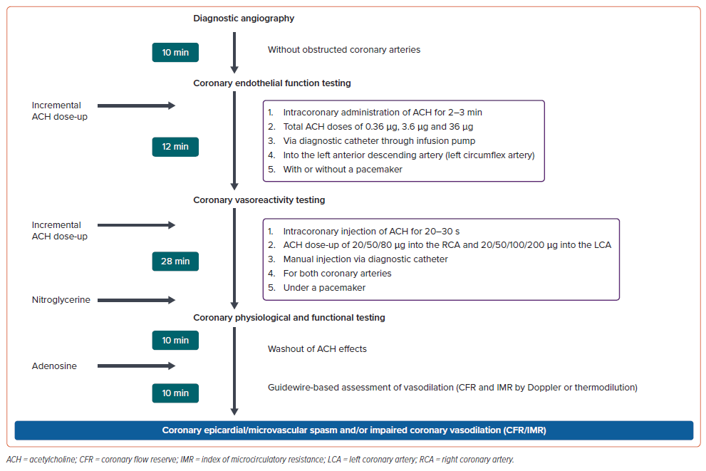
Global guidelines for the diagnosis of epicardial spasm, coronary microvascular spasm and coronary microvascular dysfunction are necessary, as are unified diagnostic strategies for use in the cardiac catheterisation laboratory. As shown in Figure 2, the proposed ideal diagnostic interventional procedures may be cumbersome and time-consuming. A procedure time of approximately 1 hour is necessary to diagnose coronary spasm and coronary endothelial dysfunction. Furthermore, radiation exposure and contrast medium due to repetitive coronary angiography are serious problems.

Figure 2: Proposed Interventional Diagnostic Strategy Protocol

Article image

The cardiologist may perform culling vasoreactivity testing of ACH if patients have no coronary constriction after the injection of low-dose ACH. However, if cardiologists around the world perform this diagnostic interventional procedure carefully in the cardiac catheterisation laboratory, they may reach a satisfactory diagnosis for each patient.

Global guidelines should be established to unify the diagnosis of epicardial spasm, coronary microvascular spasm and coronary microvascular dysfunction, and the COVADIS, ESC, AHA/ACC, ACI-SEC (Interventional Cardiology Association of the Spanish Society of Cardiology) and JCS guidelines.

Conclusion

Vasoreactivity testing with ACH has two diagnostic functions: to identify coronary endothelial function and to investigate coronary microvascular spasm and epicardial spasm. However, each physician uses a different modified ACH protocol to diagnose epicardial spasm, coronary microvascular spasm and coronary endothelial dysfunction. Unified ACH testing for coronary spasm and endothelial function is, therefore, essential for cardiologists.

Click here to view Supplementary Material.

References

  1. Ludmer PL, Selwyn AP, Shook TL, et al. Paradoxical vasoconstriction induced by acetylcholine in atherosclerotic coronary arteries. N Engl J Med 1986;315:1046–51. 
    Crossref | PubMed
  2. ENCORE Investigators. Effect of nifedipine and cerivastatin on coronary endothelial function in patients with coronary artery disease. The ENCORE I study (Evaluation of Nifedipine and Cerivastatin On Recovery of coronary Endothelial function). Circulation 2003;107:422–8. 
    Crossref | PubMed
  3. Yasue H, Horio Y, Nakamura N, et al. Induction of coronary artery spasm by acetylcholine in patients with variant angina: possible role of the parasympathetic nervous system in the pathogenesis of coronary artery spasm. Circulation 1986;74:955–63. 
    Crossref | PubMed
  4. Furchgott RF, Zawadzki JV. The obligatory role of endothelial cells in the relaxation of arterial smooth muscle by acetylcholine. Nature 1980;288:373–6. 
    Crossref | PubMed
  5. Bairey Merz CN, Kelsey SF, Pepine CJ, et al. The Women’s Ischemia Syndrome Evaluation (WISE) study: protocol design, methodology and feasibility report. J Am Coll Cardiol 1999;33:1453–61. 
    Crossref | PubMed
  6. Wei J, Mehta PK, Johnson BD, et al. Safety of coronary reactivity testing in women with no obstructive coronary artery disease: results from the NHLBI-sponsored WISE (Women’s Ischemia Syndrome Evaluation) study. JACC Cardiovasc Interv 2012;5:646–53. 
    Crossref | PubMed
  7. Ford TJ, Stanley B, Good R, et al. Stratified medical therapy using invasive coronary function testing in angina: the CorMicA Trial. J Am Coll Cardiol 2018;72:2841–55. 
    Crossref | PubMed
  8. Widmer RJ, Samuels B, Samady H, et al. The functional assessment of patients with non-obstructive coronary artery disease: expert review from an international microcirculation working group. EuroIntervention 2019;14:1694–702. 
    Crossref | PubMed
  9. Kunadian V, Chieffo A, Camici PG, et al. An EAPCI Expert Consensus Document on ischemia with non-obstructive coronary arteries in collaboration with European Society Cardiology Working Group on Coronary Pathophysiology & Microcirculation endorsed by Coronary Vasomotor Disorders International Study Group. Eur Heart J 2020;41:3504–20. 
    Crossref | PubMed
  10. Ford TJ, Ong P, Sechtem U, et al. Assessment of vascular dysfunction in patients without obstructive coronary artery disease: why, how, and when. JACC Cardiovasc Interv 2020;13:1847–64. 
    Crossref | PubMed
  11. Lee BK, Lim HS, Fearon WF, et al. Invasive evaluation of patients with angina in the absence of obstructive coronary artery disease. Circulation 2015;131:1054–60. 
    Crossref | PubMed
  12. Pargaonkar VS, Lee JH, Chow EKH, et al. Dose-response relationship between intracoronary acetylcholine and minimal lumen diameter in coronary endothelial function testing of women and men with angina and no obstructive coronary artery disease. Circ Cardiovasc Interv 2020;13:e008587. 
    Crossref | PubMed
  13. Gutierrez E, Gomez-Lara J, Escaned J, et al. Assessment of the endothelial function and spasm provocation test performed by intracoronary infusion of acetylcholine. Technical report from the ACI-SEC. REC Interv Cardiol 2021;3:286–96. 
    Crossref
  14. Egashira K, Suzuki S, Hirooka Y, et al. Impaired endothelium-dependent vasodilation of large epicardial and resistance coronary arteries in patients with essential hypertension. Different responses to acetylcholine and substance P. Hypertension 1995;25:201–6. 
    Crossref | PubMed
  15. Akiyama Y, Matoba T, Katsuki S, et al. Comparison of endothelial dysfunction in coronary arteries with bare metal and 2nd-generation drug-eluting stents. J Atheroscler Thromb 2022;29:379–92. 
    Crossref | PubMed
  16. Aziz A, Hansen HS, Sechtem U, et al. Sex-related differences in vasomotor function in patients with angina and unobstructed coronary arteries. J Am Coll Cardiol 2017;70:2349–58. 
    Crossref | PubMed
  17. Sueda S, Kohno H. The acetylcholine administration time plays the key role for provoked spasm in the spasm provocation test. J Cardiol 2017;70:141–6. 
    Crossref | PubMed
  18. Ong P, Athanasiadis A, Borgulya G, et al. Clinical usefulness, angiographic characteristics, and safety evaluation of intracoronary acetylcholine provocation testing among 921 consecutive white patients with unobstructed coronary arteries. Circulation 2014;129:1723–30. 
    Crossref | PubMed
  19. Ong P, Athanasiadis A, Hill S, et al. Coronary artery spasm as a frequent cause of acute coronary syndrome: the CASPAR (Coronary Artery Spasm in Patients with Acute Coronary Syndrome) study. J Am Coll Cardiol 2008;52:523–7. 
    Crossref | PubMed
  20. Ong P, Athanasiadis A, Sechtem U. Intracoronary acetylcholine provocation testing for assessment of coronary vasomotor disorders. J Vis Exp 2016;(114):54295. 
    Crossref | PubMed
  21. JCS Joint Working Group. Guidelines for diagnosis and treatment of patients with vasospastic angina (coronary spastic angina) (JCS 2013). Circ J 2014;78:2779–801. 
    Crossref | PubMed
  22. Sueda S, Kohno H, Miyoshi T, et al. Maximal acetylcholine dose of 200 mg into the left coronary artery as a spasm provocation tests: comparison with 100 mg of acetylcholine. Heart Vessels 2015;30:771–8. 
    Crossref | PubMed
  23. Okumura K, Yasue H, Horio Y, et al. Multivessel coronary spasm in patients with variant angina: a study with intracoronary injection of acetylcholine. Circulation 1988;77:535–42. 
    Crossref | PubMed
  24. Okumura K, Yasue H, Matsuyama K, et al. Sensitivity and specificity of intracoronary injection of acetylcholine for the induction of coronary artery spasm. J Am Coll Cardiol 1988;12:883–8. 
    Crossref | PubMed
  25. Mohri M, Koyanagi M, Egashira K, et al. Angina pectoris caused by coronary microvascular spasm. Lancet 1998;351:1165–9. 
    Crossref | PubMed
  26. Sueda S, Ochi N, Kawada H, et al. Frequency of provoked coronary vasospasm in patients undergoing coronary arteriography with spasm provocation test of acetylcholine. Am J Cardiol 1999;83:1186–90. 
    Crossref | PubMed
  27. Wakabayashi K, Suzuki H, Honda Y, et al. Provoked coronary spasm predicts adverse outcome in patients with acute myocardial infarction: a novel predictor of prognosis after acute myocardial infarction. J Am Coll Cardiol 2008;52:518–22. 
    Crossref | PubMed
  28. Ohba K, Sugiyama S, Sumida H, et al. Microvascular coronary artery spasm presents distinctive clinical features with endothelial dysfunction as nonobstructive coronary artery disease. J Am Heart Assoc 2012;1:e002485. 
    Crossref | PubMed
  29. Satoh S, Omura S, Inoue H, et al. Clinical impact of coronary artery spasm in patients with no significant coronary stenosis in acute coronary syndromes. J Cardiol 2013;61:404–9. 
    Crossref | PubMed
  30. Odaka Y, Takahashi J, Tsuburaya R, et al. Plasma concentration of serotonin is a novel biomarker for coronary microvascular dysfunction in patients with suspected angina and unobstructive coronary arteries. Eur Heart J 2017;38:489–96. 
    Crossref | PubMed
  31. Suda A, Takahashi J, Hao K, et al. Coronary functional abnormalities in patients with angina and nonobstructive coronary artery disease. J Am Coll Cardiol 2019;74:2350–60. 
    Crossref | PubMed
  32. Sueda S, Sakaue T. Intracoronary acetylcholine testing among 746 consecutive Japanese patients with angina-like chest pain and unobstructed coronary artery disease. Eur Heart J Open 2022;2:oeab012. 
    Crossref | PubMed
  33. Pristipino C, Beltrame JF, Finocchiaro ML, et al. Major racial differences in coronary constrictor response between Japanese and Caucasians with recent myocardial infarction. Circulation 2000;101:102–8. 
    Crossref | PubMed
  34. Kim JW, Park CG, Suh SY, et al. Comparison of frequency of coronary spasm in Korean patients with versus without myocardial bridging. Am J Cardiol 2007;100:1083–6. 
    Crossref | PubMed
  35. Ong P, Athanasiadis A, Borgulys G, et al. High prevalence of a pathological response to acetylcholine testing in patients with stable angina pectoris and unobstructed coronary arteries. The ACOVA study (Abnormal Coronary Vasomotion in Patients with Stable Angina and Unobstructed Coronary Arteries). J Am Coll Cardiol 2012;59:655–62. 
    Crossref | PubMed
  36. Schoenenberger AW, Felber S, Gujer S, et al. Invasive findings in patients with angina equivalent symptoms but no coronary artery disease; results from the heart quest cohort study. Int J Cardiol 2013;167:168–73. 
    Crossref | PubMed
  37. Montone RA, Niccoli G, Fracassi F, et al. Patients with acute myocardial infarction and non-obstructive coronary arteries: safety and prognostic relevance of invasive coronary provocative tests. Eur Heart J 2018;39:91–8. 
    Crossref | PubMed
  38. Seitz A, Gardezy J, Pirozzolo G, et al. Long-term follow-up in patients with stable angina and unobstructed coronary arteries undergoing intracoronary acetylcholine testing. JACC Cardiovasc Interv 2020;13:1865–76. 
    Crossref | PubMed
  39. Jansen TPJ, Elias-Smale SE, van den Oord S, et al. Sex differences in coronary function test results in patients with angina and nonobstructive disease. Front Cardiovasc Med 2021;8:750071. 
    Crossref
  40. Sun H, Mohri M, Shimokawa H, et al. Coronary microvascular spasm causes myocardial ischemia in patients with vasospastic angina. J Am Coll Cardiol 2002;39:847–51. 
    Crossref | PubMed
  41. Beltrame JF, Crea F, Kaski JC, et al. International standardization of diagnostic criteria for vasospastic angina. Eur Heart J 2017;38:2565–8. 
    Crossref | PubMed
  42. Ong P, Camici PG, Beltrame JF, et al. International standardization of diagnostic criteria for microvascular angina. Int J Cardiol 2018;250:16–20. 
    Crossref | PubMed
  43. Takahashi J, Suda A, Nishiyama K, et al. Pathophysiology and diagnosis of coronary functional abnormalities. Eur Cardiol 2021;16:e30. 
    Crossref | PubMed
  44. Ford TJ, Berry C. How to diagnose and manage angina without obstructive coronary artery disease: lessons from the British Heart Foundation CorMicA trial. Interv Cardiol 2019;14:76–82. 
    Crossref | PubMed
  45. Luscher TF, Pieper M, Tendera M, et al. A randomized placebo-controlled study on the effect of nifedipine on coronary endothelial function and plaque formation in patients with coronary artery disease: the ENCORE II study. Eur Heart J 2009;30:1590–7. 
    Crossref | PubMed
  46. Knuuti J, Wijns W, Saraste A, et al. ESC guidelines for the diagnosis and management of chronic coronary syndromes of the European Society of Cardiology (ESC). Eur Heart J 2020;41:407–77. 
    Crossref | PubMed
  47. Anderson JL, Adams CD, Antman EM, et al. 2012 ACCF/AHA focused update incorporated into the ACCF/AHA 2007 guidelines for the management of patients with unstable angina/non-ST-elevation myocardial infarction: a report of the American College of Cardiology Foundation/American Heart Association Task Force on Practice Guidelines. Circulation 2013;127:e663–828. 
    Crossref | PubMed